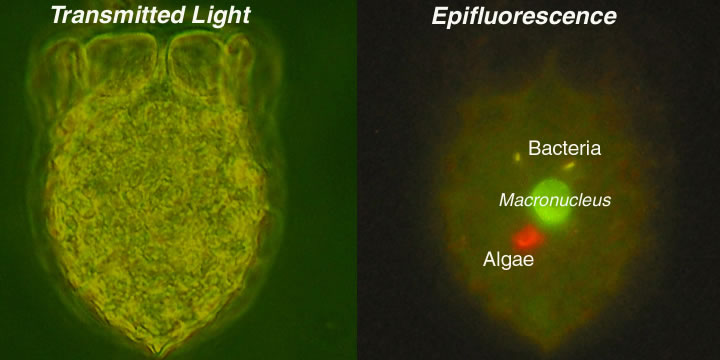
Dictyocysta mitra : the last supper
Back to Dolan Page
This is a an image of the tintinnid ciliate Dicytocysta mitra from the Bay of Villefranche. On the left is a standard light microscope image. On the right is the same specimen viewed using epifluorescence. The image on the right shows the light emitted from different cell contents due to the UV excitation. The yellow spots are photosynthetic bacteria, Synechococcus, the red spot is chlorophyll fluorescence from a microscopic algae. The tintinnid had ingested the bacteria and the algae, shortly before being removed from the Bay and preserved with gluteraldehyde. The gluteraldehyde makes the macronucleus of the cell green when subjected to UV excitement.