SO_DYFAMED Time Series - 1991-> ... |
|
JC. MARTY : head of mission and project leader |
|
PIGMENTS HPLC : JC. MARTY |
METHOD | PUBLICATIONS | FIGURES
(get
data set in excel file format : ![]() )
)
Method for HPLP pigments analysis |
||||||||||||
|
Water
for pigment analysis (2 liters) was filtrated on 25 mm Whatman GF/F
glass fiber filters. The filters were frozen and analyzed by HPLC
within 3 months. Filters were ground and sonicated in 3-ml methanol
(HPLC grade) under dim light conditions. The method used until 1993
was derived from that of Mantoura and Llewellyn (1983). The general
procedure for HPLC pigment analysis, identification and
quantification has been described (Claustre et
al., 1994 a,b). With the separation system used (RP-C18), a
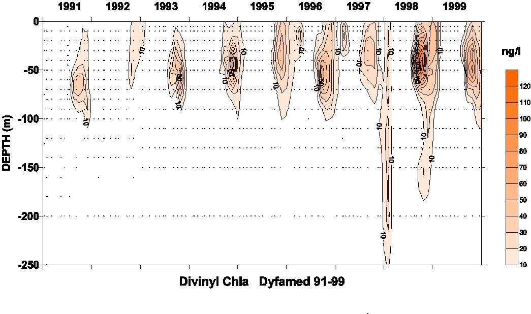
partial resolution of divinyl-chlorophyll a
(DV Chl a) from
chlorophyll a (Chl a) has been
achieved. For samples from 1994 and later, the separation method
used (RP-C8) is described in Vidussi et
al. (1996), and the resolution between DV Chl a
and Chl a has been
complete. The
continuity of the set of data was obtained by the utilization of an
internal standard (b-apocarotenal) added to each sample in the extraction solvent. The
possible effect of the change of analytical method was tested by
analyzing the same samples with the two procedures. The agreement
between the two methods was good (+- 5%) of the same order than the
agreement between 2 analyses of the same sample using the same
method. A special attention was given to the quantification of DV
Chl a, which is fully
resolved from Chl a in the
second part of the experiment. Although partial, the separation of
these two compounds in the first phase of our study was sufficient
for a good matching of the data from the two methods (equivalent to
other pigments) except for low concentrations of DV Chl a
(below 5 ng l-1) not detected in the first method. Results
are reported in terms of Chl a,
divinyl-chlorophyll a (DV
Chl a) and Total Chl a (TChl a
= Chl a + DV Chl a). Chlorophyll
b (Chl b)
and divinyl-chlorophyll b (DV
Chl b) not resolved with the first separation method, and partially
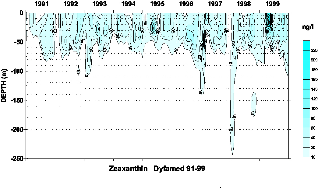
resolved by the new one, are presented together as TChl b. Lutein and zeaxanthin were partially resolved using the method of
Vidussi et al. (1996), but
data are presented as the sum of the two compounds. The lutein was
only occasionally detected and always at very low levels with
respect to zeaxanthin. Then the couple lutein-zeaxanthin can be
considered as essentially zeaxanthin. Chlorophylls
and carotenoids were detected and quantified by absorbance at 440
nm. Identification of pigments was performed by comparison of
on-line collected absorption spectra with those of a library of
spectra established from standards and reference cultures obtained
from the Villefranche sur mer culture collection. The standard
carotenoids used for the calibration of the HPLC [peridinin (peri),
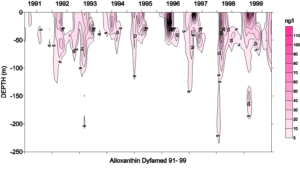
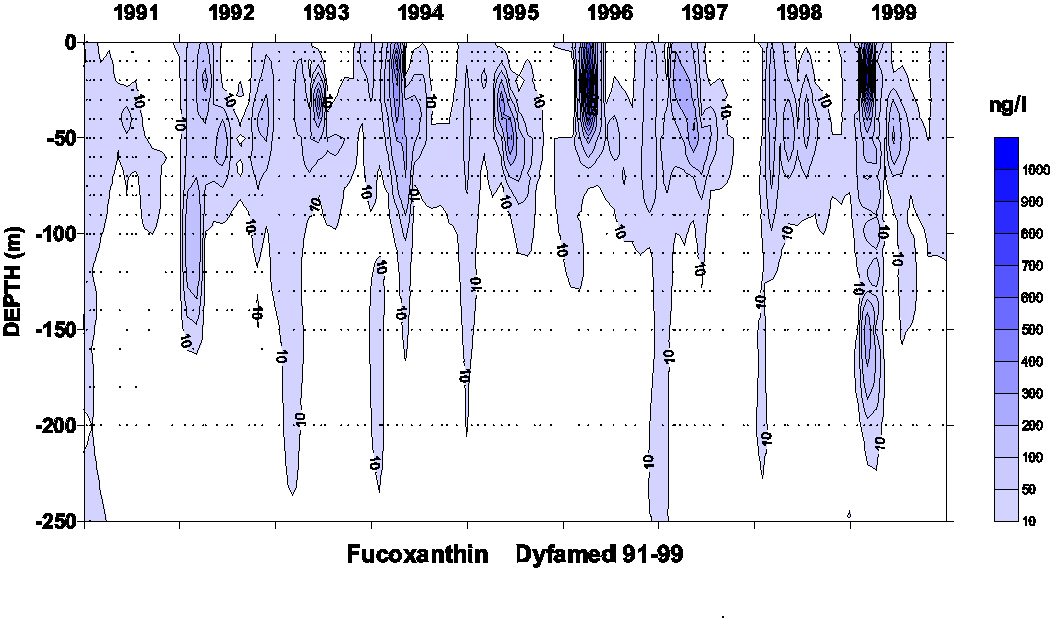
alloxanthin (allo), fucoxanthin (fuco), zeaxanthin (zea),
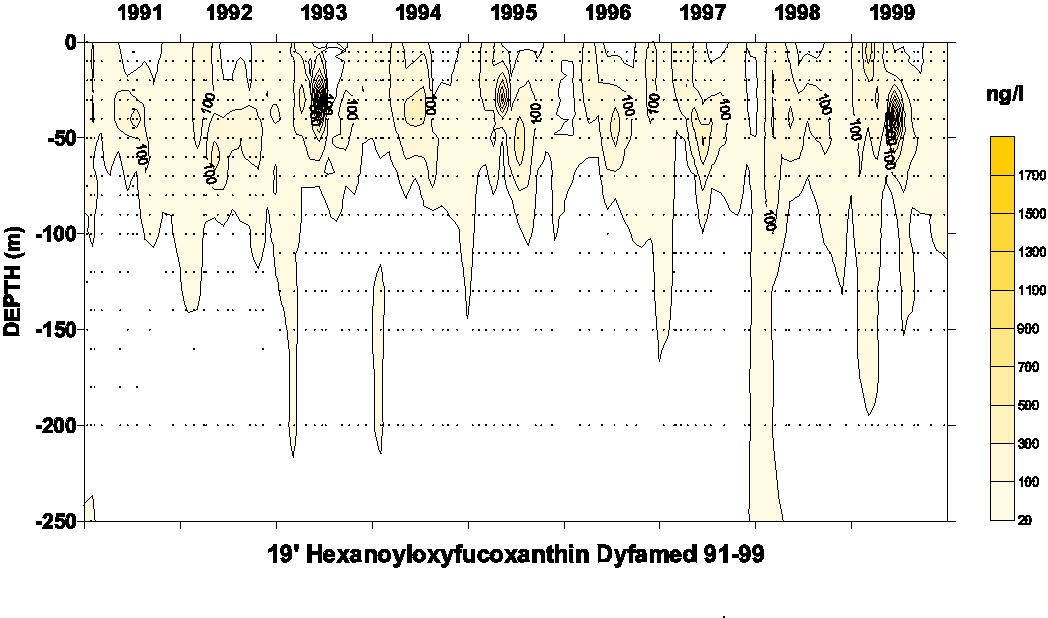
19’-hexanoyloxyfucoxanthin (19’HF), 19’-butanoyloxyfucoxanthin
(19’BF)] were provided by R. Bidigare as part of a JGOFS
intercalibration exercise. Chlorophyll a and chlorophyll b were from
Sigma Chemical Co. Diode Array detection was achieved on selected
samples until 1993 (Waters 991) and on all samples since 1994 (HP
1100). A
range of phytoplankton pigments has been detected, in order to
characterize different phytoplankton groups. A recent review of
taxonomic pigments can be found in Jeffrey (1997).
Divinyl-chlorophyll a is
the typical marker of prochlorophytes whereas Chl a
is the universal descriptor of other phytoplankton taxa. Fucoxanthin
(Fuco) characterizes diatoms and peridinin (peri) dinoflagellates.
Nano- and pico-flagellates containing chlorophyll c
are characterized by 19'-hexanoyloxyfucoxanthin (19'HF,
prymnesiophytes) and by 19'-butanoyloxyfucoxanthin (19'BF,
chrysophytes and pelagophytes). Zeaxanthin (Zea) is the marker of
cyanobacteria but it is also present in prochlorophytes. All
data are available through the DYFAMED Observatory data base http://www.obs-vlfr.fr/jgofs2/sodyf/home.htm
. Contour maps were obtained using Surfer program (Golden software Inc.) and Kriging method.
|
||||||||||||
Bibliography |
||||||||||||
|
Claustre,
H., Kerhervé, P., Marty, J.C., Prieur, L., Videau, C., Hecq, J.H.,
1994. Phytoplankton
dynamics associated with a geostrophic front: ecological and
biogeochemical implications. Journal of Marine Research 52, 711-742. Claustre,
H., Kerhervé, P., Marty, J.C., Prieur, L., 1994.
Phytoplankton
photoadaptation in relation to some frontal physical processes.
Journal of Marine Systems 5, 251-265. Jeffrey, S.W., 1997. Application of pigment methods to oceanography. In: Jeffrey, S.W., Mantoura, R.F.C., Wright, S.W. (Eds.), Phytoplankton pigments in oceanography. UNESCO, Paris, pp. 127-178. Mantoura,
R.F.C., Llewellyn, C.A., 1983. The
rapid determination of algal chlorophyll and carotenoid pigments and
their breakdown products in natural waters by reverse-phase
high-performance liquid chromatography. Analytica Chimica Acta 151,
293-314. Vidussi, F., Claustre, H., Bustillos-Guzman, J., Cailliau, C., Marty, J.C., 1996. Determination of chlorophylls and carotenoids of marine phytoplankton : separation of chlorophyll a from divinyl-chlorophyll a and zeaxanthin from lutein. Journal of Plankton Research 18, 2377-2382.
|
||||||||||||
FIGURES |
||||||||||||
|